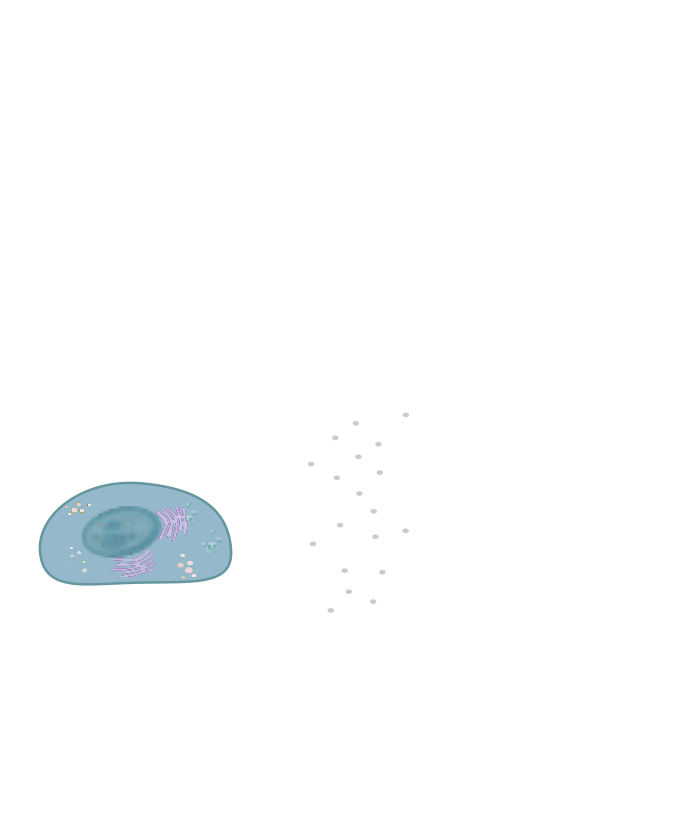
Our vision is to understand biology one molecule at a time. We strive to provide a dynamic and quantitative understanding in structural and cell biology and to utilize this information to control aberrant biological function. We approach this formidable challenge with an eclectic mix of quantitative single particle microscopy techniques and machine learning analysis.
The main objective of my group is to augment our understanding on the molecular mechanisms that
underlie and control vital cellular functions. We approach this challenge by deciphering the dynamic
interplay between the function and spatiotemporal localization of biomolecules (virus, dug
nanocarrier, oligonucleotides or protein assemblies) and how this correlate to cellular and
organismal response. We utilize an eclectic mix of single particle techniques that promises to shed
light on the interplays between the behaviour (dynamics, function and localisation) of biomolecules
and high throughput single particle screening methodologies to decipher oligonucleotide interactions
with membranes.
Recognizing that 4D imaging generates terabytes of data sets, that are hard to be quantitatively
evaluated by current semi-manual analysis, we have developed toolboxes based on machine learning to
rapidly and reliably, analyze the wealth of novel microscopy data we, and others, produce. Our
all-inclusive softwares for windows and macs, offer accelerate by 5 orders of magnitude transition
from raw data to quantitative analysis. These combined methodologies bridge 4D imaging with
sophisticate image analysis required for delving into the era of 4D cell and tissue imaging.
Our
Funding
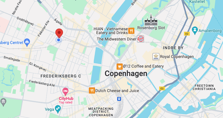
Thorvaldsensvej 40
Frederiksberg 1871